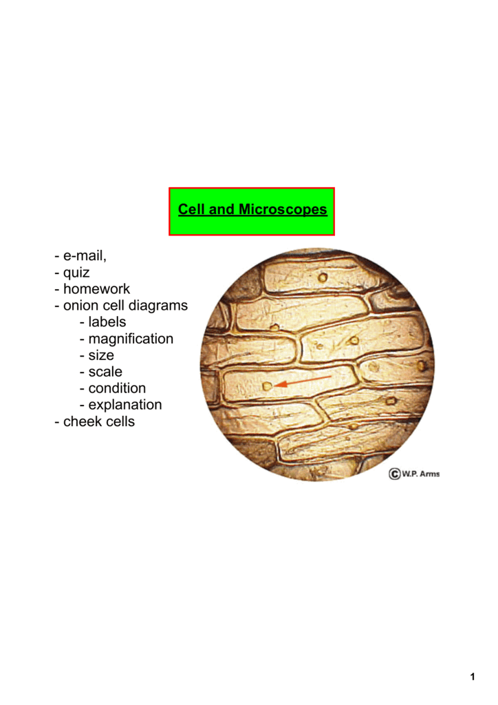
38 onion cells under microscope with labels
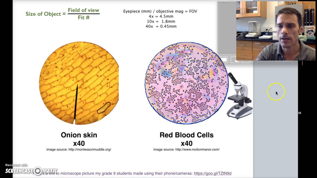
Cambridge International AS and A Level Biology Coursebook … Cambridge International AS and A Level Biology Coursebook Fourth Edition Plant Cell Under Microscope Labeled 40X : Young Root 2 Of Broad Bean ... Cells and viewing them under the microscope. A small square of a red onion skin (membrane) was observed under a microscope at high power (x40) magnification. (iv) describe how you applied the stain. They must draw and label the nucleus, cell membrane set up your microscope, place the onion root slide on the stage and focus on low (40x) power.
ONION CELLS VIDEO - YouTube Video shows how to make a wet mount slide to view onion cells under the microscope.
Onion cells under microscope with labels
Onion Cells Under a Microscope (100x-2500x) - YouTube In this video you will see onion cells under a microscope (100x-2500x) as is, without any coloring. To observe the onion cells the thin membrane is used. It... GeM | Bidding Advanced search for Ongoing Bids. Search by Bid / RA Details; Search by Ministry / Organization; Bid / RA Number Observing Onion Cells Under The Microscope Afterwards, carefully mount the prepared and stained onion cell slide onto the microscope stage. Make sure that the cover slip is perfectly aligned with the microscope slide, and that any excess stain has been wiped off. Secure the slide on the stage using the stage clips.
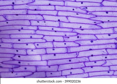
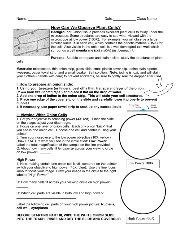
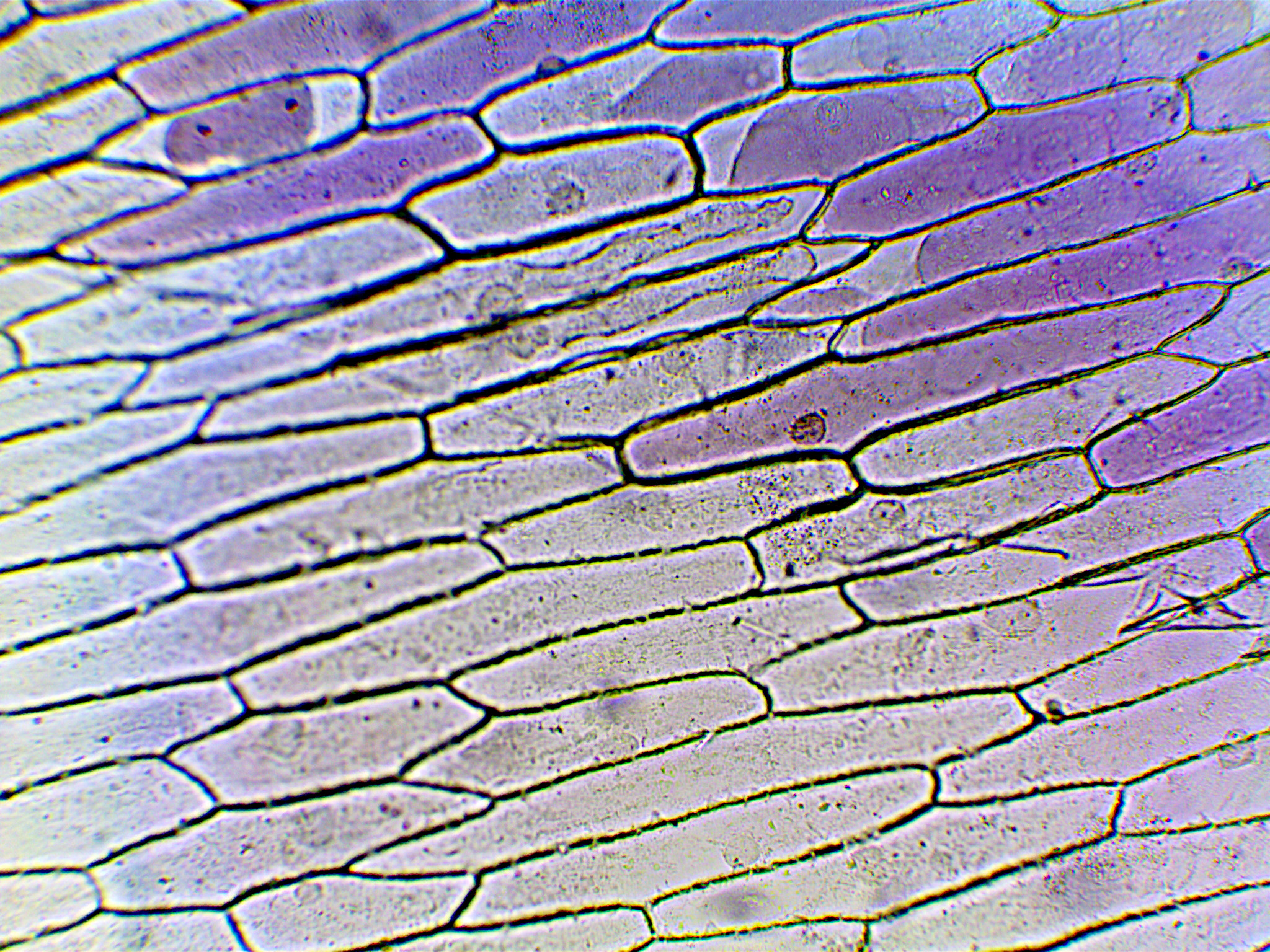
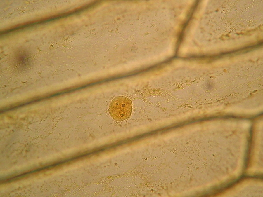
Onion cells under microscope with labels. DOC The Onion Cell Lab - chsd.us Onion tissue provides excellent cells to study under the microscope. The main cell structures are easy to see when viewed with the microscope at medium power. For example, you will observe a large circular . nucleus. in each cell, which contains the genetic material for the cell. In each nucleus, are round bodies called . nucleoli Natural Sciences Grade 9 - Grade 7-9 Workbooks Robert Hooke (1635 - 1703). Robert Hooke was the first cytologist to identify cells under his microscope in 1665. He decided to call the microscopic shapes that he saw in a slice of cork "cells" because the shapes reminded him of the cells (rooms) that the monks in the nearby monastery lived in.Robert Hooke was the first to use the term 'cell' when he studied thin slices … Leaf Cell Under Microscope Labeled 2 päivää sitten · leaf cell under microscope labeled. Leaf Cell Under Microscope Labeled. w7m, ur3, knj4, ... The following diagram shows cells of onion peel label class ... - Vedantu 115.2k + views. Hint: The diagrams mentioned above are the internal structure of an onion peel and human cheek cells. In order to label them, we need to understand its anatomy and know about various structures present in it. Onion peel is an example of a plant cell whereas a human cheek cell is an example of an animal cell. Complete answer:
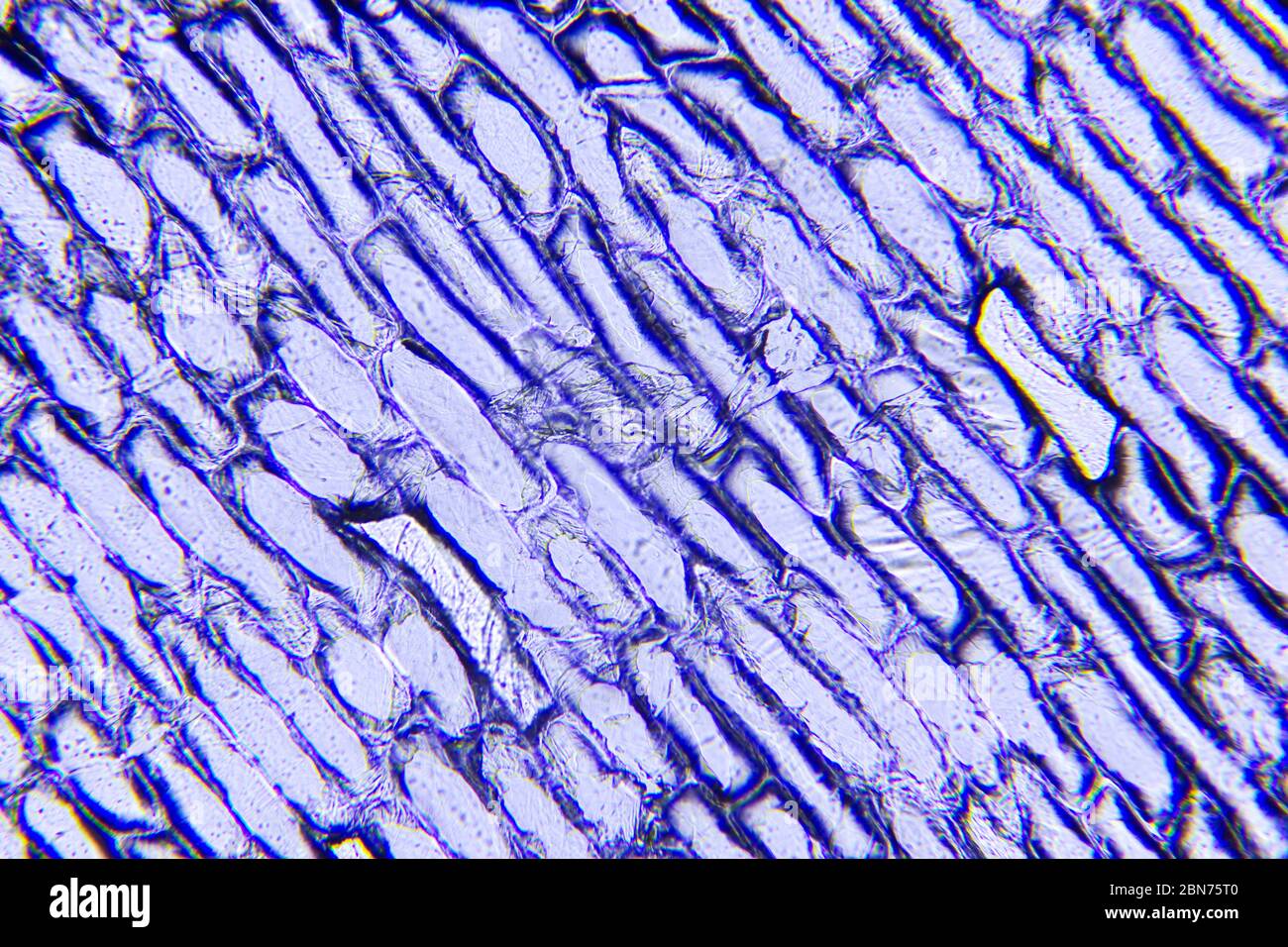
Microscope Cell Lab: Cheek, Onion, Zebrina - SchoolWorkHelper The first lab exercise was observing animal cells, in this case, my cheek cells. The second lab exercise was observing plant cells, in this case, onion epidermis. The third lab exercise was observing chloroplasts and biological crystals, in this case, a thin section from the Zebrina plant. The first thing that was done in this lab exercise was ... Onion Cells Microscope Stock Photos and Images - Alamy Onion cells under the microscope. Garden onion, Bulb Onion, Common Onion (Allium cepa), cell tissue of a garden onion with dyed chromosomes, light microscopy, x 120, Germany. Onion Cells under the Microscope. Onion skin cells under the microscope, horizontal field of view is about 0.61 mm. Detailed view of the cells of a red onion as seen ... PDF Onion Cells - Investigation - Exploring Nature 5. Observe the onion tissue under the microscope at 4x, 10x and 40x with lots of light (open diaphragm). Then slowly close the diaphragm while observing the image to find the best light for seeing cellular details. 6. Draw a section of onion skin cells at 10x magnification. Then switch to 40x and draw one cell and label it. Questions: 1. How to observe onion cells under a microscope? - JacAnswers How to observe onion cells under a microscope? Gently lay a microscopic cover slip on the membrane and press it down gently using a needle to remove air bubbles. Touch a blotting paper on one side of the slide to drain excess iodine/water solution, Place the slide on the microscope stage under low power to observe.
(PDF) Taiz & Zeiger- Plant Physiology | Munish K Bansal Taiz & Zeiger- Plant Physiology Onion Epidermal Cell Labeled Diagram - schematron.org Draw a labelled diagram of an onion epidermal cell seen under the microscope. ( 4 marks) e The onion epidermal cells are not green in colour because they lack. The epidermal cells of onions provide a protective layer against viruses and fungi that may harm the sensitive tissues. News Archives | Hollywood.com Travel through time by exploring Hollywood.com's entertainment news archives, with 30+ years of entertainment news content. PDF Onion Cell Lab - somewaresinmaine.com Research Biology Onion Cell Lab page 1 of 3 Onion Cell Lab After you have completed the rest of this lab come back to this cover page DRAW & LABEL AN ONION CELL WITH ALL THE PARTS / ORGANELLES YOU OBSERVE UNDER 40X. Purpose: To observe and identify major plant cell structures and to relate the structure of the cell to its function. Materials: 1 ...
Onion Leaf Cell Under Microscope - Micropedia
Onion Root Tip Mitosis - Stages, Experiment and Results · Cover the sample (root tip) with a coverslip and gently press the coverslip down, then examine the slide under the microscope starting with low magnification * For this experiment, a properly prepared slide should appear light pink due to the stain to almost colorless. * Unused roots can be stored in 70 percent alcohol. Results

Onion Epidermal Cell Labeled - Top Label Maker
Cells and Reproduction - BBC Bitesize The proper name for a living thing is a living organism. A living organism can be, amongst other things, a plant or an animal.
Onion Cells High Resolution Stock Photography and Images - Alamy
The Biology Project The Biology Project, an interactive online resource for learning biology developed at The University of Arizona. The Biology Project is fun, richly illustrated, and tested on 1000s of students. It has been designed for biology students at the college and high school level, but is useful for medical students, physicians, science writers, and all types of interested people.

Living and Learning: Testing out my Microscope
DOC Plant and Animal Cells Microscope Lab - Hillsboro City Schools Students will observe onion cells under a microscope. Students will discover that their skin is made up of cells. Students will observe cheek cells under a microscope. Materials: microscope. ... Draw a diagram of one cheek cell and label the parts. (You should observe the cell membrane, nucleus, and cytoplasm.)

swifty science: onion cell lab
Onion Cell Labeled Under Microscope Search: Onion Cell Under Microscope Labeled. View the cells under the microscope using scanning power (40x magnification) to find your sample on the slide Start with the lowest power objective and work your way up , cheek cells, onion cells) under a microscope or similar instrument, and draw labelled biological diagrams to show how the cells' organelles differ Things to Do: Procedure: 1 ...

The Cells and Microorganisms Webquest
Under the Micrsocope: Onion Cell (100x - 400x) - YouTube In this "experiment" we will see onion cells under the microscope.For the experiment you will only need onion, dropper and the microscope (container and tool...

SENTHIL PRABHU SIVASAMY: Observation of Plant & Animal Cells
Microscopy, size and magnification - Microscopy, size and ... - BBC Place cells on a microscope slide. Add a drop of water or iodine (a chemical stain). Lower a coverslip onto the onion cells using forceps or a mounted needle. This needs to be done gently to...

Phases of mitosis in control and 1-day CA-treated root tip cells. Bars... | Download Scientific ...
Nano based drug delivery systems: recent developments and … 19.9.2018 · Recently, there has been enormous developments in the field of delivery systems to provide therapeutic agents or natural based active compounds to its target location for treatment of various aliments [33, 34].There are a number of drug delivery systems successfully employed in the recent times, however there are still certain challenges that need to be addresses and an …

Onion cells under microscopes | News | Wimbledon High School
Onion Root Mitosis - Microscopy-UK Onions have larger chromosomes than most plants and stain dark. The chromosomes are easily observed through a compound light microscope. The cells pictured below are located in the apical meristem of the onion root. The apical meristem is an area of a plant where cell division takes place at a rapid rate. Phases of plant cells division:

Rens blog : Science, cells
Onion Cell Lab Report.docx - Onion Cell Lab Report By Onion Cell Lab Report By : Nawaf Almalki Introduction: Many things that are viewed using a microscope, particularly cells, can appear quite transparent under the microscope. The internal parts of the cells, the organelles, are so transparent that they are often difficult to see. Biologists have developed a number of stains that help them see the cells and their organelles by adding color to ...
Labeled Onion Cell Under Microscope 40x - Micropedia
Onion Cells Under a Microscope - Requirements/Preparation/Observation Add a drop of iodine solution on the onion membrane (or methylene blue) Gently lay a microscopic cover slip on the membrane and press it down gently using a needle to remove air bubbles. Touch a blotting paper on one side of the slide to drain excess iodine/water solution, Place the slide on the microscope stage under low power to observe.
Microscope Onion Cell Labeled - Micropedia
Observing Onion Cells Under The Microscope Afterwards, carefully mount the prepared and stained onion cell slide onto the microscope stage. Make sure that the cover slip is perfectly aligned with the microscope slide, and that any excess stain has been wiped off. Secure the slide on the stage using the stage clips.
Onion Cells under Microscope
GeM | Bidding Advanced search for Ongoing Bids. Search by Bid / RA Details; Search by Ministry / Organization; Bid / RA Number

onion cells through microscope | I put my camera right up to… | Flickr
Onion Cells Under a Microscope (100x-2500x) - YouTube In this video you will see onion cells under a microscope (100x-2500x) as is, without any coloring. To observe the onion cells the thin membrane is used. It...
The inner epidermis of the onion bulb’s cataphylls (the onion skin).
Onion Cell Under Microscope 4x 10x 40x - Micropedia
Post a Comment for "38 onion cells under microscope with labels"