42 microscope diagram with labels and definitions
7th grade Science - Microscope Diagram | Quizlet The Parts of a Microscope. 12 terms. totobear PLUS. Sets found in the same folder. Science Key terms 7th grade. 13 terms. palocastillo. 7th Grade Earth Science. 9 terms. EliseC17. 7thGrade Review - Cells/Biology. 26 terms. SolizScience TEACHER. 7th grade Science, Cell theory. 8 terms. Super1412. Other sets by this creator. Inorganic Chemistry 4th edition, Catherine Housecroft Enter the email address you signed up with and we'll email you a reset link.
Types of Microscopes: Definition, Working Principle, Diagram ... A simple microscope is defined as the type of microscope that uses a single lens for the magnification of the sample. A simple microscope is a convex lens with a small focal length. The magnifying power of the simple microscope is given as: m = 1 + D F Where, D is the least distinct vision F is the focal length of the convex lens
Microscope diagram with labels and definitions
Compound Microscope: Definition, Diagram, Parts, Uses, Working ... - BYJUS A microscope with a high resolution and uses two sets of lenses providing a 2-dimensional image of the sample. The term compound refers to the usage of more than one lens in the microscope. Also, the compound microscope is one of the types of optical microscopes. The other type of optical microscope is a simple microscope. Diagram (Parts labelled), Principle and Uses - Microscope Wiki The three structural components include: 1. Head - This is the upper part of the microscope that houses the optical parts 2. Arm - This part connects the head with the base and provides stability to the microscope. Arm is used to carry the microscope around 3. Base - Base is on which the microscope rests and the base houses the illuminator that lights up the specimens Laboratory procedures for diagnosis of anthrax, and isolation and ... In the case of Member States with limited resources and unable to operate at BSL3, it is pertinent to remember that B. anthracis is not highly infectious, and that humans are moderately resistant (see section 4.2.1). For diagnostic test purposes, therefore, good laboratory practice (Table 12) at all times is the important factor in carrying out the necessary tests safely. Large numbers of the ...
Microscope diagram with labels and definitions. Microscope, Microscope Parts, Labeled Diagram, and Functions Microscope, Microscope Parts, Labeled Diagram, and Functions What is Microscope? A microscope is a laboratory instrument used to examine objects that are too small to be seen by the naked eye. It is derived from Ancient Greek words and composed of mikrós, "small" and skopeîn,"to look" or "see". Parts of Stereo Microscope (Dissecting microscope) - labeled diagram ... Labeled part diagram of a stereo microscope Major structural parts of a stereo microscope Optical components of a stereo microscope - definition and function Eyepieces Eyepiece tube Diopter adjustment ring Interpupillary Adjustment Objective Lenses Barlow lens Adjustment Knobs Light sources Stage plate Stage chips Histopathological Image Analysis: A Review - PMC Histopathology is the study of the signs of the disease using the microscopic examination of a biopsy or surgical specimen that is processed and fixed onto glass slides. To visualize different components of the tissue under a microscope, the sections are dyed with one or more stains. Compound Microscope Parts, Diagram Definition, Application, Working ... Compound Microscope Parts, Diagram Definition, Application, Working Principle. ... For future reference, adhesive labels are stuck to the base and sides of the microscope. (iii) Use. After calibrating the eyepiece scales for all objective lenses, the microscope can be used to measure the dimensions and morphology of cells and sub-cellular ...
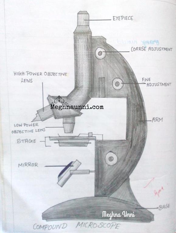
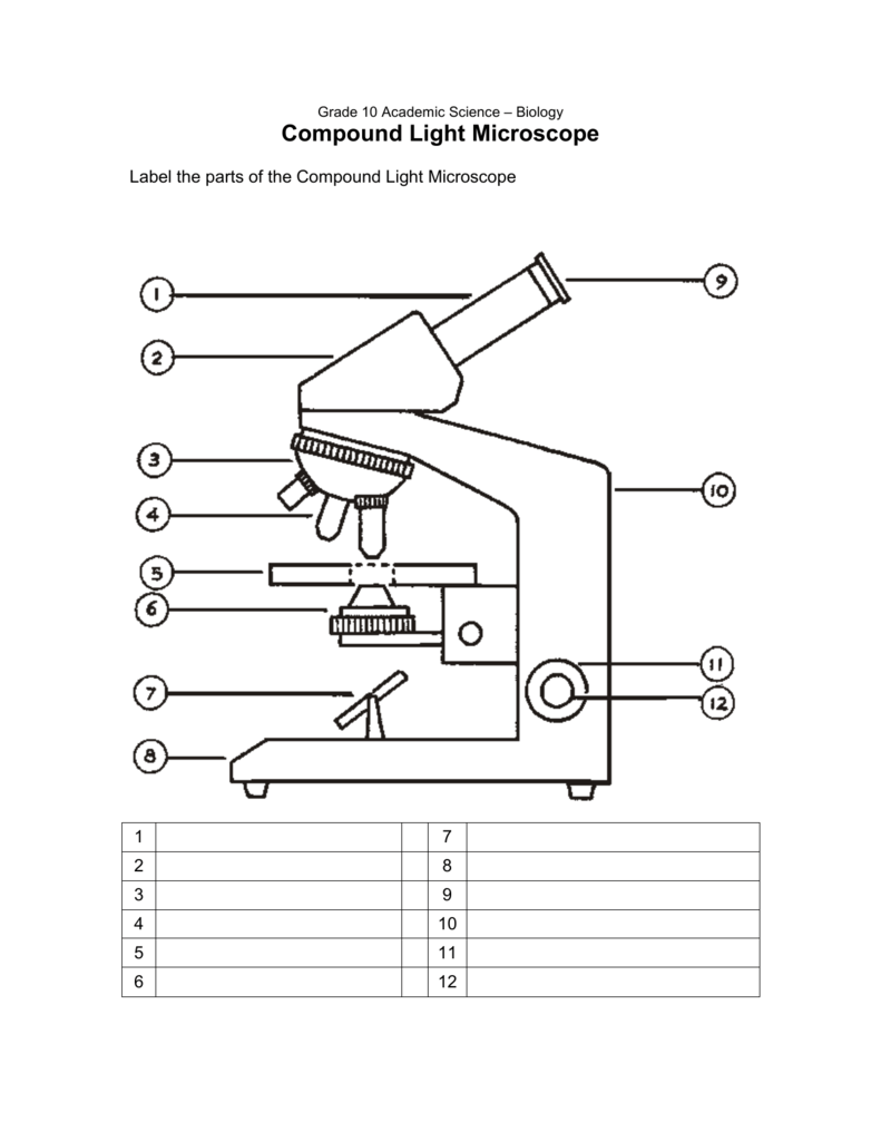
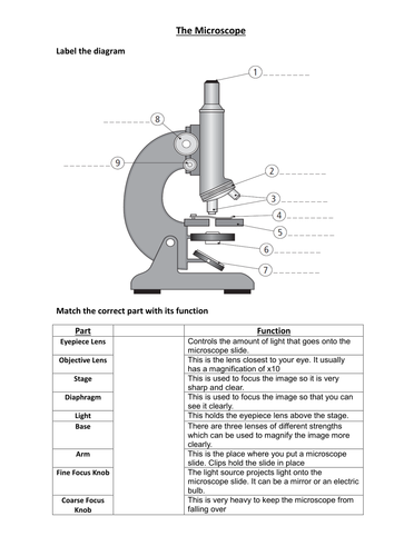
Fountain Essays - Your grades could look better! All our academic papers are written from scratch. All our clients are privileged to have all their academic papers written from scratch. These papers are also written according to your lecturer’s instructions and thus minimizing any chances of plagiarism. PDF Parts of the Light Microscope - Science Spot B. NOSEPIECE microscope when carried Holds the HIGH- and LOW- power objective LENSES; can be rotated to change MAGNIFICATION. Power = 10 x 4 = 40 Power = 10 x 10 = 100 Power = 10 x 40 = 400 What happens as the power of magnification increases? Simple Microscope - Parts, Functions, Diagram and Labelling Parts of the optical parts are as follows: Mirror - A simple microscope has a plano-convex mirror and its primary function is to focus the surrounding light on the object being examined. Lens - The biconvex lens is placed above the stage and its function is to magnify the size of the object being examined. E-Learning – AOAC India Bright Field Microscope, Dark Field Microscope, Phase Contrast Microscope, Fluorescence Microscope, Confocal microscopy, Scanning and Transmission Electron Microscope and applications: 17th June 2019, 2019 11.30-12.30 pm: Watch Video: 50 : Nuclear magnetic resonance (NMR) – Part 1 DR. CHANDRASHEKHAR MR. RAGHAV MAVINKURVE, BRUKER
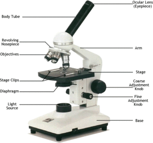
Compound Microscope- Definition, Labeled Diagram, Principle, Parts, Uses The term "compound" in compound microscopes refers to the microscope having more than one lens. Devised with a system of combination of lenses, a compound microscope consists of two optical parts, namely the objective lens and the ocular lens. Working Principle of the Compound Microscope Label Microscope Diagram - EnchantedLearning.com low-power objective - a small lens with low magnifying power. mirror (or light source) - this directs light upwards onto the slide. revolving nosepiece - the rotating device that holds the objectives (lenses). stage - the platform on which a slide is placed. stage clips - metal clips that hold a slide securely onto the stage. Parts of the Microscope with Labeling (also Free Printouts) Parts of the Microscope with Labeling (also Free Printouts) A microscope is one of the invaluable tools in the laboratory setting. It is used to observe things that cannot be seen by the naked eye. Table of Contents 1. Eyepiece 2. Body tube/Head 3. Turret/Nose piece 4. Objective lenses 5. Knobs (fine and coarse) 6. Stage and stage clips 7. Aperture Metalloid - Wikipedia A metalloid is a type of chemical element which has a preponderance of properties in between, or that are a mixture of, those of metals and nonmetals.There is no standard definition of a metalloid and no complete agreement on which elements are metalloids. Despite the lack of specificity, the term remains in use in the literature of chemistry.
Labelled Microscope Diagram Gcse - Micropedia
CODEX multiplexed tissue imaging with DNA-conjugated … 02-07-2021 · This protocol describes co-detection by indexing, a highly multiplexed imaging technology that uses DNA-conjugated antibodies to image up to 60 markers in formalin-fixed, paraffin-embedded and ...
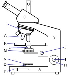
Diagram Of A Microscope With Labels - Drivenhelios
Fountain Essays - Your grades could look better! Professional academic writers. Our global writing staff includes experienced ENL & ESL academic writers in a variety of disciplines. This lets us find the most appropriate writer for any type of assignment.

Microscope Diaphragm Definition - Micropedia
Simple Microscope - Diagram (Parts labelled), Principle, Formula and Uses A simple microscope consists of Optical parts Mechanical parts Labeled Diagram of simple microscope parts Optical parts The optical parts of a simple microscope include Lens Mirror Eyepiece Lens A simple microscope uses biconvex lens to magnify the image of a specimen under focus.

All Saints Online
Parts of a microscope with functions and labeled diagram Microscope Definition Microscopes are instruments that are used in science laboratories to visualize very minute objects such as cells, and microorganisms, giving a contrasting image that is magnified. Microscopes are made up of lenses for magnification, each with its own magnification powers.
Labelled Pictures Of Human Skin : Skin Diagram With Detailed Illustrations And Clear Labels ...
16 Parts of a Compound Microscope: Diagrams and Video Body of the Microscope In compound microscopes with two eye pieces there are prisms contained in the body that will also split the beam of light to enable you to view the image through both eye pieces. 2. Arm The arm of the microscope is another structural piece. The arm connects the base of the microscope to the head/body of the microscope.

Animal Cell- Definition, Structure, Parts, Functions and Diagram
Unmasking – Part 2 | The Vineyard of the Saker Jun 01, 2022 · None of the old “isms” or ideological labels are useful anymore. Therefore, all of the theories of economics should be regarded with the same approach as archaeology, which can help understand how the current runaway insane systems were originally developed, but no longer are able to explain the absurdities that have finally resulted from ...
Meghna Unni's Blog: Meghna's Paintings, Bharathanatyam, Writings, Stories, Projects, Crafts
Histopathological Image Analysis: A Review - PMC From the Voronoi diagram, two more graphs of interest can be constructed: the Delaunay triangulation, which is created by connecting points that share an edge in the Voronoi diagram, and the minimum spanning tree, which is the series of lines that spans the set of points such that the Euclidean sum of the lengths of the lines is smaller than any other spanning tree.

PPT - Types of Organisms PowerPoint Presentation, free download - ID:2471768
Fuzzy concept - Wikipedia A fuzzy concept is a concept of which the boundaries of application can vary considerably according to context or conditions, instead of being fixed once and for all. This means the concept is vague in some way, lacking a fixed, precise meaning, without however being unclear or meaningless altogether. It has a definite meaning, which can be made more precise only …
zHomeschooler's Resources: November 2010
Microscope Parts and Functions With Labeled Diagram and Functions How ... First, the purpose of a microscope is to magnify a small object or to magnify the fine details of a larger object in order to examine minute specimens that cannot be seen by the naked eye. Here are the important compound microscope parts... Eyepiece: The lens the viewer looks through to see the specimen.

10.13.08: Histology - Bone Formation and Remodeling
University of South Carolina on Instagram: “Do you know a ... Oct 13, 2020 · I’m a real and legit sugar momma and here for all babies progress that is why they call me sugarmomma progress I will bless my babies with $2000 as a first payment and $1000 as a weekly allowance every Thursday and each start today and get paid 💚
![How to Use a Microscope: Lesson for Kids - Science Class [2021] | Study.com](https://study.com/cimages/multimages/16/labeledmicroscopeimage.jpg)
How to Use a Microscope: Lesson for Kids - Science Class [2021] | Study.com
Compound Microscope Parts - Labeled Diagram and their Functions - Rs ... The eyepiece (or ocular lens) is the lens part at the top of a microscope that the viewer looks through. The standard eyepiece has a magnification of 10x. You may exchange with an optional eyepiece ranging from 5x - 30x. [In this figure] The structure inside an eyepiece. The current design of the eyepiece is no longer a single convex lens.
33 Microscope Diagram To Label - Labels Database 2020
Labeling the Parts of the Microscope | Microscope World Resources Labeling the Parts of the Microscope This activity has been designed for use in homes and schools. Each microscope layout (both blank and the version with answers) are available as PDF downloads. You can view a more in-depth review of each part of the microscope here. Download the Label the Parts of the Microscope PDF printable version here.

34 How To Label Parts Of A Picture In Word - Label Ideas 2020
The Parts of a Microscope (Labeled) Printable - TeacherVision The Parts of a Microscope (Labeled) Printable. Download. Add to Favorites. Share. This diagram labels and explains the function of each part of a microscope. Use this printable as a handout or transparency to help prepare students for working with laboratory equipment. Grade:

Label Microscope Diagram - ClipArt Best
Parts of the Microscope Label and Definition Diagram | Quizlet Controls amount of light entering the body tube Light Source sends light upward through the diaphragm Ocular Lens (Eye Piece) Holds magnifying lens used for viewing Arm supports body tube Stage Holds slide on flat surface Coarse Adjustment Knob Moves Body Tube up and down for focusing Fine Adjustment Knob Sharpens image by moving body tube slightly
Microscope Labelled Diagram Gcse - Micropedia
Cambridge International AS and A Level Biology Coursebook … Cambridge International AS and A Level Biology Coursebook Fourth Edition
Post a Comment for "42 microscope diagram with labels and definitions"