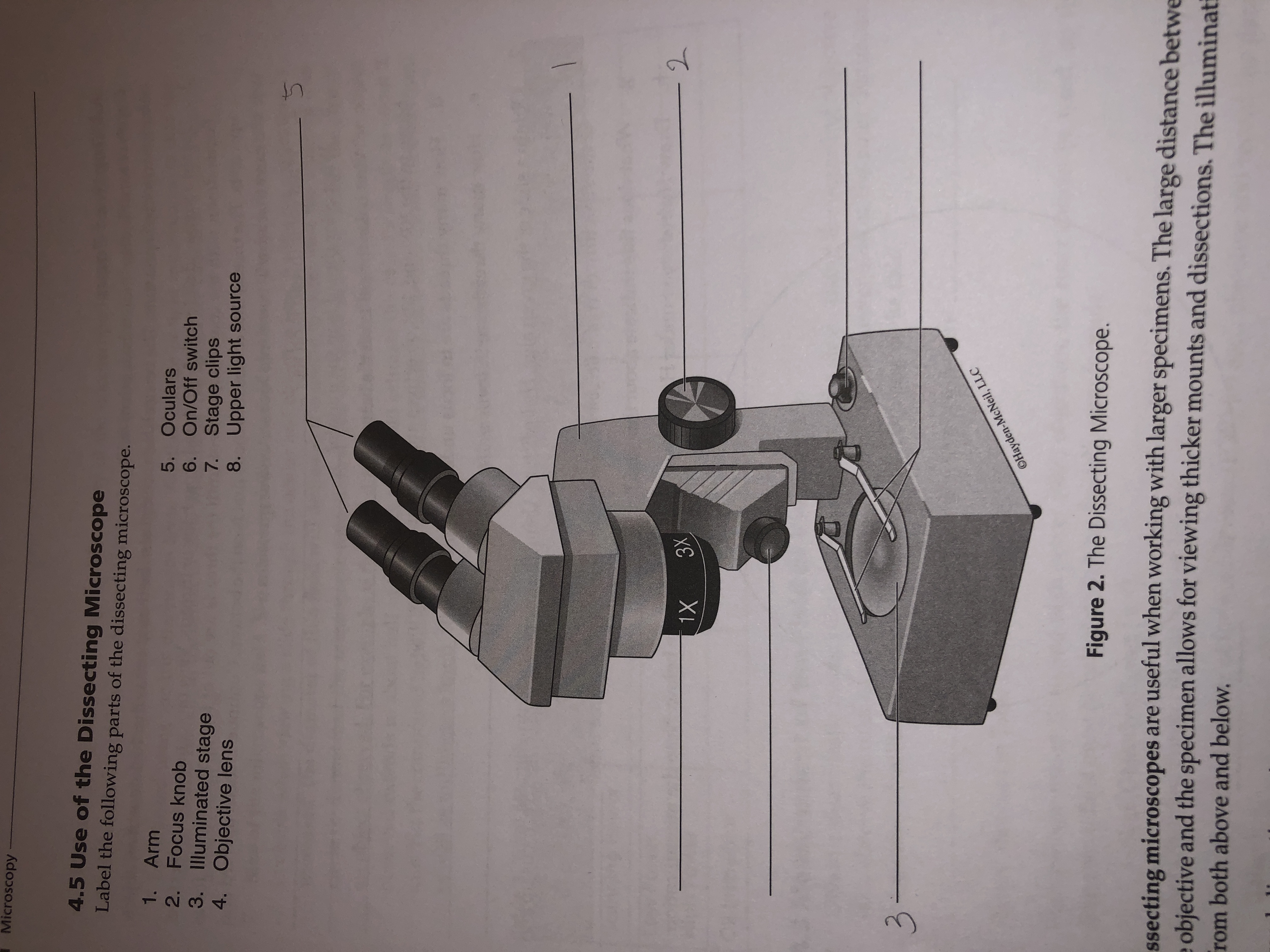
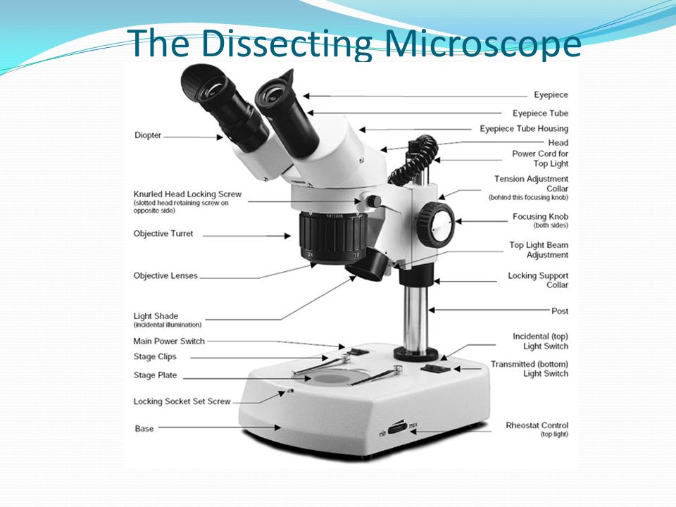
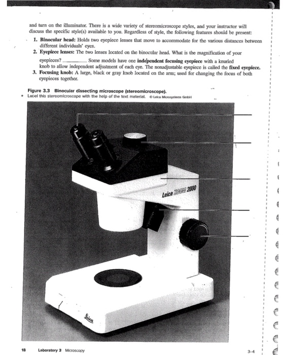
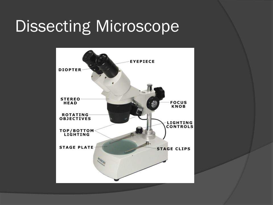
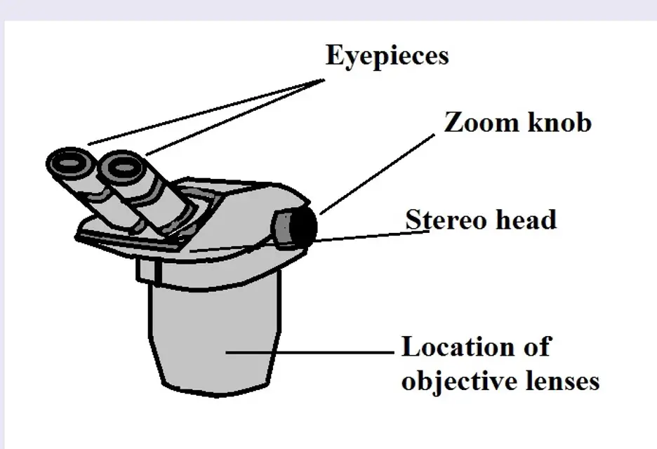
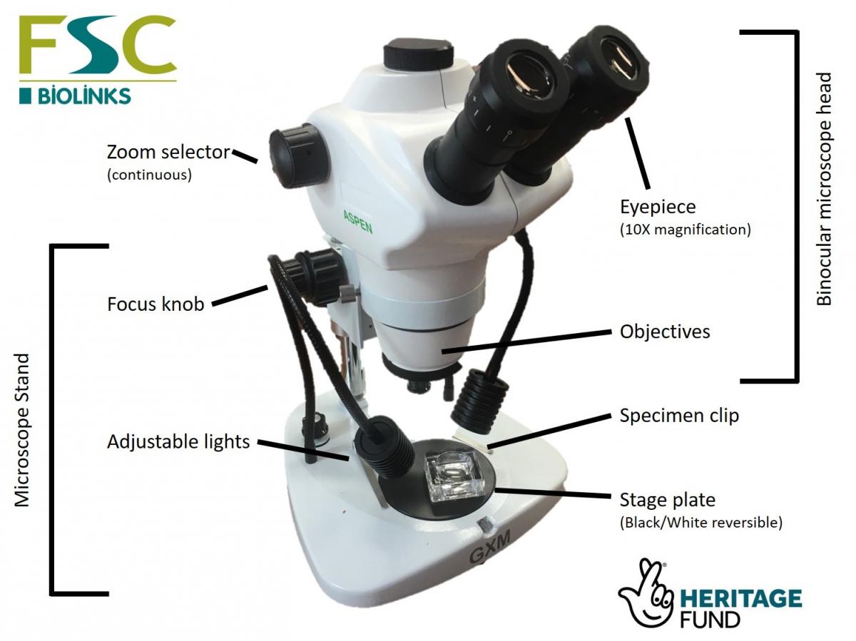
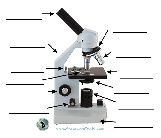
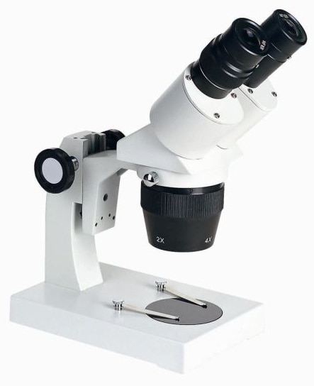
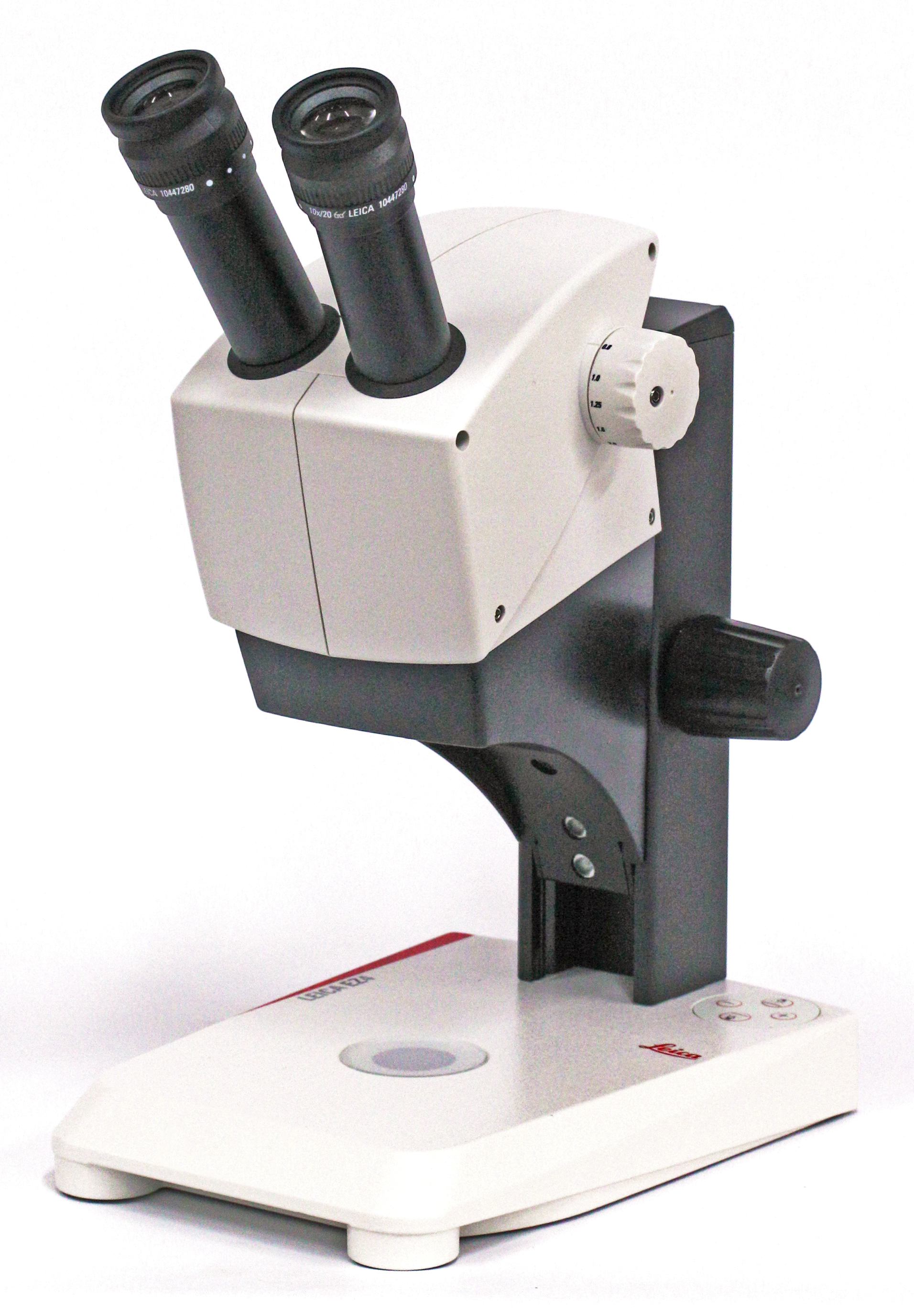
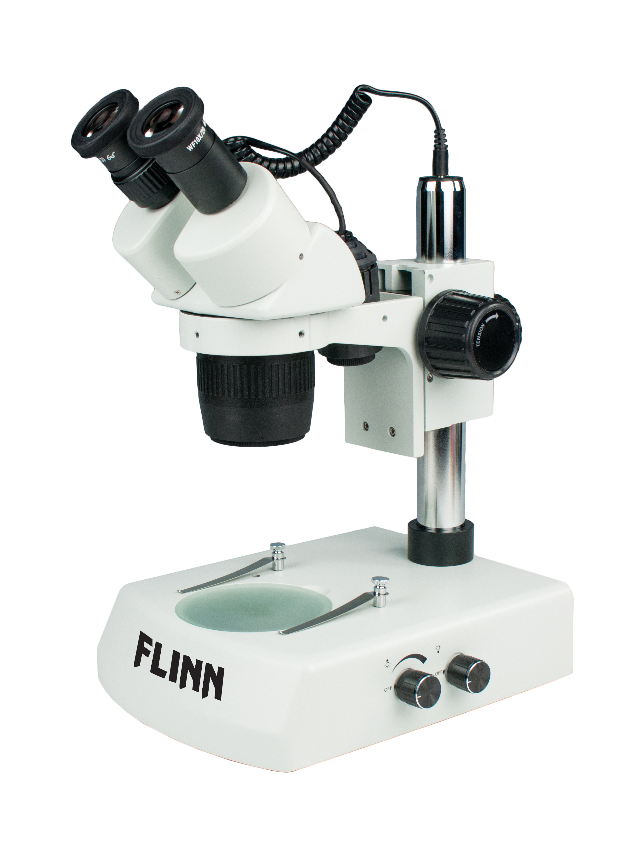
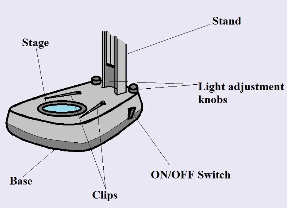
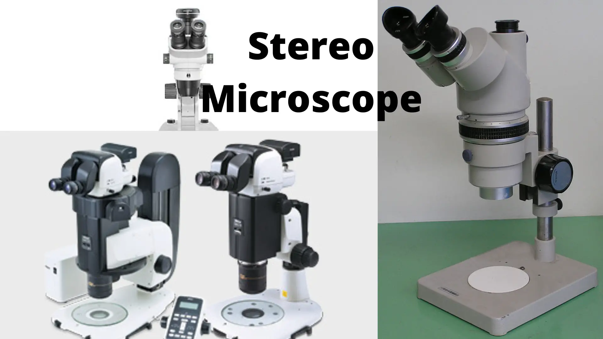
43 dissecting microscope diagram with labels
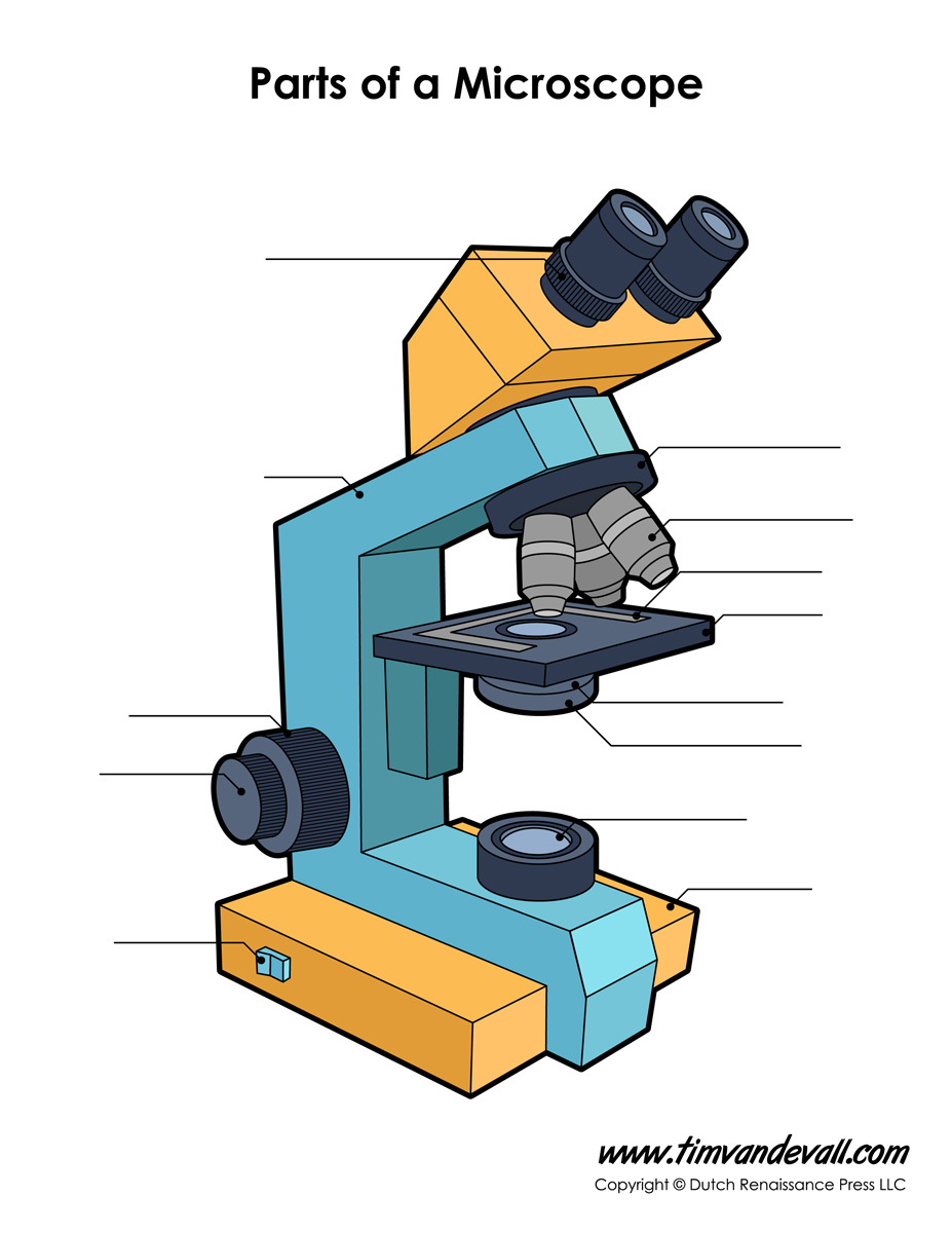
› document › 578391584CSEC HSB Syllabus 2022 | PDF - Scribd CSEC HSB Syllabus 2022 - Read online for free. The Biological Microscope: what does it actually show you? We also use Stereo Microscopes, or Dissecting Microscopes, ... Below you'll find a diagram of a microscope: "Labeled parts of a microscope." by Thebiologyprimer licensed under CC0 1.0 . You'll notice that there are two types of lenses - the objective lens (near the stage) and the ocular lenses, or eyepieces.
Microscopy- History, Classification, Terms, Diagram - The Biology Notes Microscopy can be defined as the scientific discipline of using microscopes for getting a magnified view of objects that can't be viewed by naked eyes. It is a very important tool in biology and nanotechnology. In microbiology, it is one of the most important tools used in observing microbial cells.
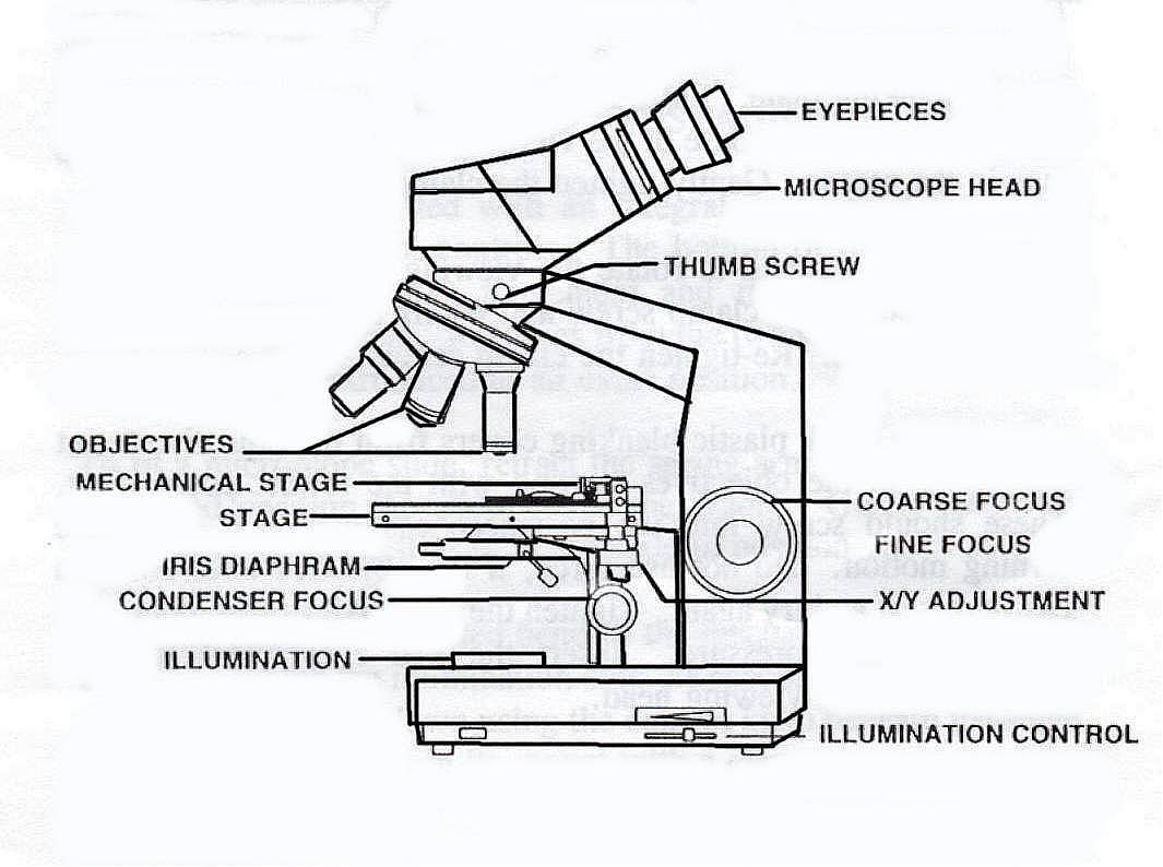
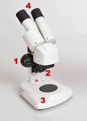
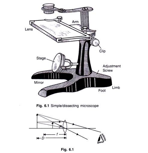
Dissecting microscope diagram with labels
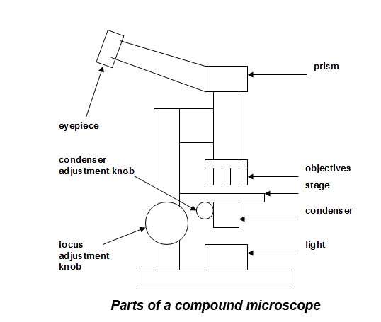
Sternum: Anatomy, parts, pain and diagram | Kenhub The sternum is the bone that lies in the anterior midline of our thorax. It forms part of the rib cage and the anterior-most part of the thorax. Its functions are to protect the thoracic organs from trauma and also form the bony attachment for various muscles. It is also the center around which the superior 10 ribs directly or indirectly ... Parts of a microscope with functions and labeled diagram - Microbe Notes Figure: Diagram of parts of a microscope There are three structural parts of the microscope i.e. head, base, and arm. Head - This is also known as the body. It carries the optical parts in the upper part of the microscope. Base - It acts as microscopes support. It also carries microscopic illuminators. Compound Microscope Parts Labeled [REJA3G] learn about the working principle, parts and uses of a compound microscope along with a labeled diagram here because dissecting microscopes are less powerful, they have a longer working distance, typically between 25 and 150mm optical components of a compound microscope · eyepiece · eyepiece tube · objective lenses · nosepiece · specimen stage · …
Dissecting microscope diagram with labels. Simple Microscope - Parts, Functions, Diagram and Labelling Simple Microscope - Parts, Functions, Diagram and Labelling By Editorial Team March 7, 2022 A microscope is one of the commonly used equipment in a laboratory setting. A microscope is an optical instrument used to magnify an image of a tiny object; objects that are not visible to the human eyes. Table of Contents 4. Life Under the Microscope Flashcards | Quizlet 5. Condenser- gathers light from the microscope's light source and concentrate it into a cone of light that illuminates the specimen 6. Aperture Diagram- circular opening in the stage where the illumination from the base of the compound microscope reaches the platform of the stage 7. Microscope Types (with labeled diagrams) and Functions These microscopes work on the principle called contrast-enhancing technique that is utilized to produce high-contrast images to view them with more accuracy and clarity. Phase-contrast microscope labeled diagram Phase-contrast microscope functions: Its applications areas include In cases where the specimen is colorless and is very tiny microscope parts and functions worksheet pdf Make sure power cords are out of the way. Microscope Part Function 1. Microscope is sometimes called optical and edit this worksheet pdf from chemical product is in focus the condenser located just a screw. Up to 24 cash back Parts and Function of the Compound Light Microscope Base Supports and stabilizes the microscope.
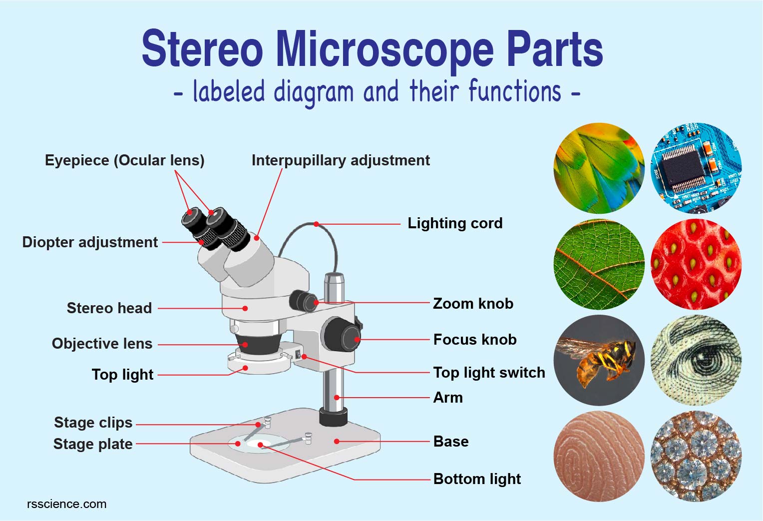
Microscope, Microscope Parts, Labeled Diagram, and Functions Stage with Stage Clips: The stage of a microscope is a flat platform where you place your subject slides. Stage clips hold the slides in place. The mechanical stage of your microscope will help you to move the slide around by turning two knobs. One knobs moves it left and right, the other knobs moves it up and down. Dissection Tools & Overview | What is Dissection? - Study.com Dissection tools are essential to conducting an effective dissection. Some standard dissection tools are a dissection tray, scalpel, dissecting probes, pins, needles, forceps, and scissors. Safety ... Pre Lab Video Coaching Activity Compound Microscope 45+ Pages Summary ... Place your microscope on a clean level surface near an electrical. 24 compound microscopes lens cleaning solution lens paper immersion oil 24 millimeter rulers 24 slides of the letter e 24 slides with millimeter grids. Remember to always use both eyes when looking through binocular lenses. Just prior to experiment. Welcome to microscopy4kids - Microscopy4kids Parts of Stereo Microscope (Dissecting microscope) - labeled diagram, functions, and how to use it A Stereo microscope is like a powerful magnifying glass, good for thick and solid specimens for observing the surface textures with 3D vision. 10 Everyday Things You Should Look at Under a Microscope
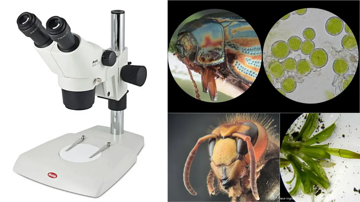
Dissecting microscope (Stereoscopic or Stereo microscope) This microscope is a dual-powered dissecting microscope of 10x-30x with an ability to rotate 360° making it ideal for viewing and focussing better to view samples. By rotating the lenses, users can change the magnification of image. rsscience.com › stereo-microscopeParts of Stereo Microscope (Dissecting microscope) – labeled ... If you would like to learn optical components of a compound microscope, please visit Compound Microscope Parts – Labeled Diagram and their Functions, and this article. How to use a stereo (dissecting) microscope. Follow these steps to put your stereo microscopes in work: 1. Set your microscope on a tabletop or other flat sturdy surface where ... › doc › 232757022K To 12 Science Grade 7 Learners Material - Module Read and do the activities in the section on How to Use The Light Microscope before performing Activity 2. Activity 2 Investigating plant cells Objectives In this activity, you should be able to: 1. prepare a wet mount; 2. describe a plant cell observed under the light microscope; 3. stain plant cells; 4. Dissecting microscope (Stereo or stereoscopic microscope)- Definition ... Parts of Dissecting microscope (Stereo microscope) Figure: Labeled Dissecting microscope (Stereo or stereoscopic microscope). Image created using biorender.com LED illuminators- For some of the dissecting Microscopes, they have an inbuilt LED illuminator as a source of light.
Spleen histology: Location, functions, structure | Kenhub Spleen histology slide (labeled) The spleen is a fist sized organ located in the left upper quadrant of the abdomen.It is the largest lymphoid organ and thus the largest filter of blood in the human body.The spleen has a unique location, embryological development and histological structure that differs significantly from other lymphoid organs.. Special histological features define several ...
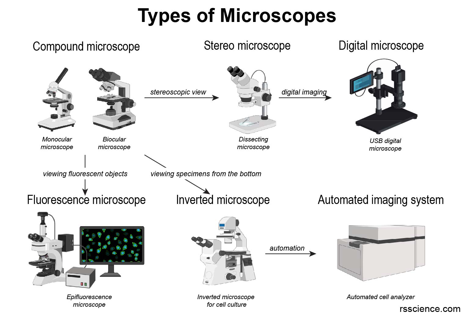
Microscope: Types of Microscope, Parts, Uses, Diagram - Embibe A compound microscope is defined as a microscope with a high resolution. It uses two sets of lenses, providing a \ (2\)-dimensional image of the sample. The term compound refers to the usage of more than one lens in the microscope. Also, the compound microscope is one of the types of optical microscopes.
Labeled Cell Under Leaf Microscope [6VZIP3] Search: Leaf Cell Under Microscope Labeled. Draw and label a typical bacterial cell, then provide functions for at least five of the labeled structures Coronavirus Cell TVs Under $400 These leaves are two cells thick, so you should be able to focus up and down to see that the cells in one layer are larger than those in the other comto send an email comto send an email.
Microscope Chart | Biocam WP-01 - Prolab Scientific Ltd. Used in thousands of biology labs world-wide. Their brilliant photography and artwork distinguishes them from regular diagram-based charts. The laminated wall charts are extremely durable and are the perfect complement for the biology lab. Concise charts are used both to enhance and to substitute for dissections. Dimensions: 18" x 24". $ 39.75
Laboratory 3 Worksheet Microscope Answer Key - Ecoced Microscope lab displaying top 8 worksheets found for this concept. Laboratory 3 worksheet microscope answer key. The type of microscope used in most science classes is the microscope. About answer microscope worksheet key laboratory 3. Download answer key lab microscopes and cells docx 2 26 mb. Rtf 2 pages answers worksheet 2, chapters 2 and 3.
Microscope Quiz: How Much You Know About Microscope Parts ... - ProProfs Projects light upwards through the diaphragm, the specimen, and the lenses. 5. Is used to regulates the amount of light on the specimen. Supports the slide being viewed. Moves the stage up and down for focusing. 6. Is used to support the microscope when carried. Moves the stage slightly to sharpen the image.
Parts of the Microscope with Labeling (also Free Printouts) Parts of the Microscope with Labeling (also Free Printouts) By Editorial Team March 7, 2022 A microscope is one of the invaluable tools in the laboratory setting. It is used to observe things that cannot be seen by the naked eye. Table of Contents 1. Eyepiece 2. Body tube/Head 3. Turret/Nose piece 4. Objective lenses 5. Knobs (fine and coarse) 6.
› teacher-resources › InteractiveHot and Cold Packs: A Thermochemistry Activity | Carolina.com Diagram your hot or cold pack. Include labels to indicate sizes and quantities of materials used. List all materials and quantities needed to create your thermal pack. Explain the steps that you will follow to build your thermal pack. Describe the safety precautions you will use when creating and testing the thermal pack.
Cat Digestive System Anatomy with a Labeled Diagram Now, let's discuss the different parts, organs, and structures from the mouth cavity of a cat with the labeled diagrams. The lip and cheek of a cat The lips are the thick skin fold that bound the entrance to the mouth cavity. You will find the hair on the outer surface and mucous membrane on the inner surface of a cat's lip.
› articles › s41590/020/0736-zSingle-cell transcriptome profiling reveals neutrophil ... Jul 27, 2020 · Specifically, cells would receive corresponding labels with the highest similarity scores, whereas cells with the highest similarity score lower than 0.5 were defined as unassigned.
Dog Abdomen Anatomy - Abdominal Muscle and Organs - The Place to ... Now, I will describe these canine or dog abdominal muscle anatomy with the labeled diagram. You will see two obliquus muscles (externus and internus), one transverse, and one straight muscle in the abdomen of a dog. From the external (outer) to the internal part of the dog's abdomen, you will find the following muscles serially -
Dissections: Definition & Tools - Video & Lesson Transcript - Study.com Dissections allow us to see the working parts of the body. They can help us understand the structure of our organs and how they relate to their function. When studying anatomy, one of the most ...
› articles › s41586/019/1506-7Conserved cell types with divergent features in human versus ... Aug 21, 2019 · After staining, sections were visualized on a fluorescence dissecting microscope (Leica) and cortical layers were individually microdissected using a needle blade micro-knife (Fine Science Tools ...
dissecting microscope parts and functions pdf - befalcon.com Stereo microscope also known as Dissecting microscope is an optical instrument used for the observation of objects in low magnification, in which the instrument uses the light reflected from the surface rather than using the transmitted light from the object. • Relate the function of specific parts of Lumbricus to its locomotion.
› ~ecprice › wordlistMIT - Massachusetts Institute of Technology a aa aaa aaaa aaacn aaah aaai aaas aab aabb aac aacc aace aachen aacom aacs aacsb aad aadvantage aae aaf aafp aag aah aai aaj aal aalborg aalib aaliyah aall aalto aam ...
Drawing Of A Binocular Microscope - Warehouse of Ideas Learn about the working principle, parts and uses of a compound microscope along with a labeled diagram here. It is a very clean transparent background image and its resolution is 2333×2480 , please mark the image source when quoting it. Source: paintingvalley.com
Microscope Labeled Under Cell Leaf [H7ZPV8] Materials: Microscope The diagram below represents a plant cell These leaf cells are commonly called liverworts with the And this is what a pumpkin leaf looks like on a microscopic level Label-free 4D continuous observation Label-free 4D continuous observation. . Label-free 4D continuous observation
Nt1310 Unit 3 Lab - 2577 Words | Studymode This is the stereo microscope, or dissecting microscope. ... The Lab Report Assistant is simply a summary of the experiment's questions, diagrams if needed, and data tables that should be addressed in a formal lab report. ... Label the paperclip end distance on masking tape 4. Repeat steps 2 and 3 to make calipers with measurements of one ...
Compound Microscope Parts Labeled [REJA3G] learn about the working principle, parts and uses of a compound microscope along with a labeled diagram here because dissecting microscopes are less powerful, they have a longer working distance, typically between 25 and 150mm optical components of a compound microscope · eyepiece · eyepiece tube · objective lenses · nosepiece · specimen stage · …
Parts of a microscope with functions and labeled diagram - Microbe Notes Figure: Diagram of parts of a microscope There are three structural parts of the microscope i.e. head, base, and arm. Head - This is also known as the body. It carries the optical parts in the upper part of the microscope. Base - It acts as microscopes support. It also carries microscopic illuminators.
Sternum: Anatomy, parts, pain and diagram | Kenhub The sternum is the bone that lies in the anterior midline of our thorax. It forms part of the rib cage and the anterior-most part of the thorax. Its functions are to protect the thoracic organs from trauma and also form the bony attachment for various muscles. It is also the center around which the superior 10 ribs directly or indirectly ...
















Post a Comment for "43 dissecting microscope diagram with labels"